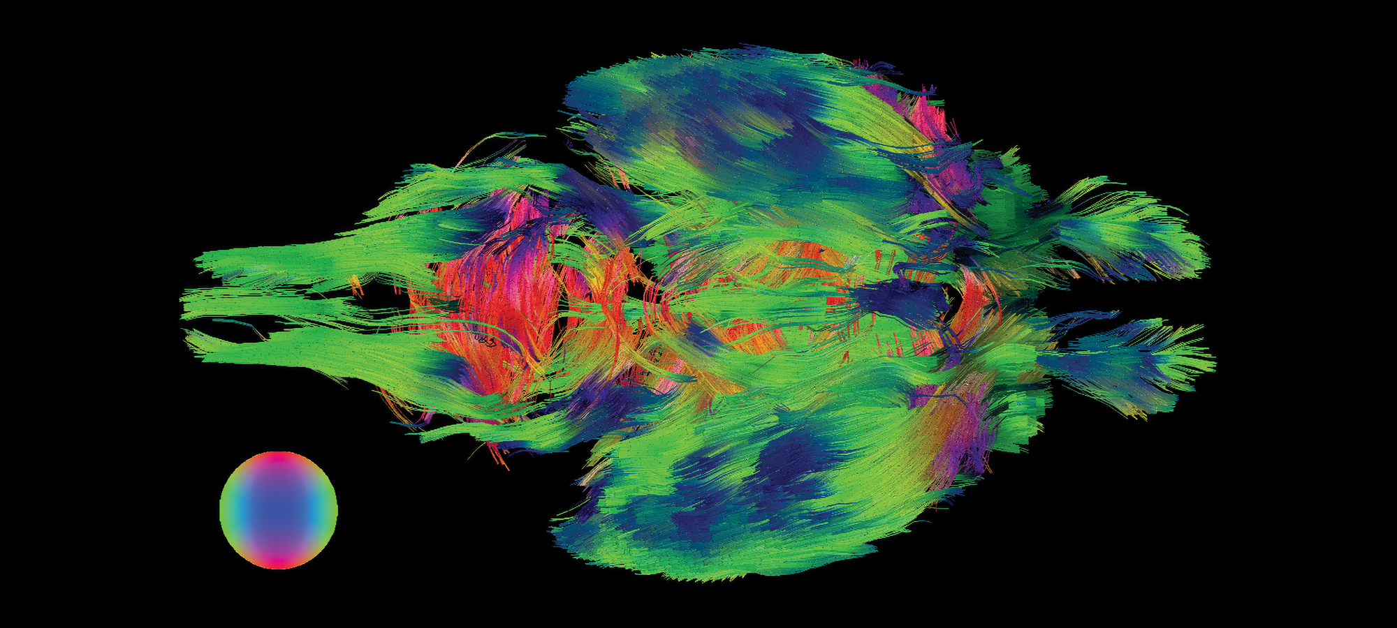
(Image courtesy Sean Foxley)
A mouse brain like you’ve never seen it before.
Itʼs a bird! Itʼs a plane! Itʼs a … mouse brain? Earlier this year a joint UChicago-Argonne team scanned an entire mouse brain at the resolution of one micron (thatʼs one millionth of a meter) using synchrotron-based X-ray tomography, a type of micro-CT scan. This approach allowed the team to bridge a gap between MRI-based visualizations (above), which show whole networks of synaptic connections but at low resolution, and electron microscopy, which shows individual neurons and synapses in detail but only a few micrometers at once. With access to all three imaging techniques, scientists can better understand the microstructure of the brain and gain new knowledge about neurological diseases. So why a mouse brain? Well, “because it fits in the microscope,” senior author Narayanan “Bobby” Kasthuri, assistant professor of neurobiology, told UChicago Medicine.
